43 eye diagram and labels
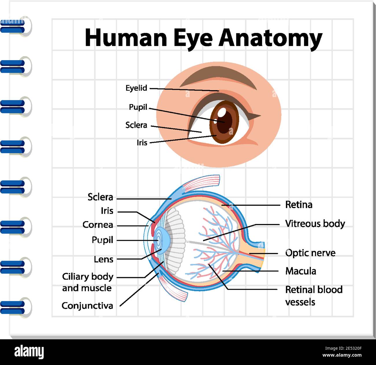
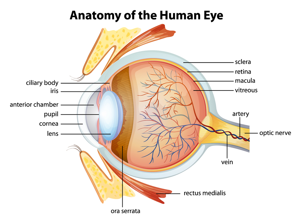
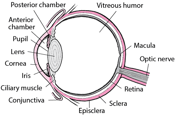
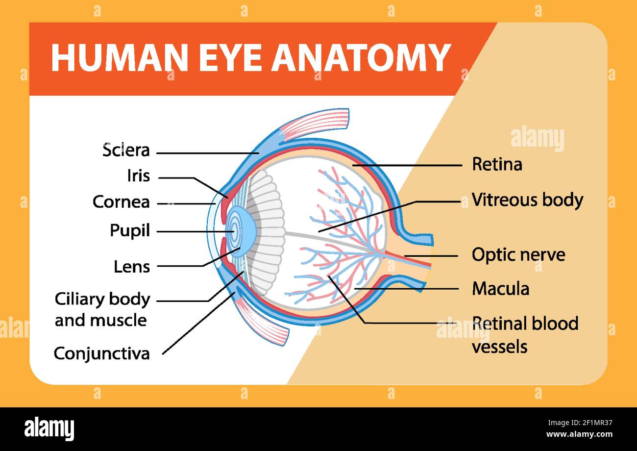
Labelled Diagram of Human Eye, Explanation and Function - VEDANTU Labeled Diagram of Human Eye, The eyes of all mammals consist of a non-image-forming photosensitive ganglion within the retina which receives light, adjusts the dimensions of the pupil, regulates the availability of melatonin hormones, and also entertains the body clock. Eye Anatomy: 16 Parts of the Eye & Their Functions - Vision Center The following are parts of the human eyes and their functions: 1. Conjunctiva, The conjunctiva is the membrane covering the sclera (white portion of your eye). The conjunctiva also covers the interior of your eyelids. Conjunctivitis, often known as pink eye, occurs when this thin membrane becomes inflamed or swollen.
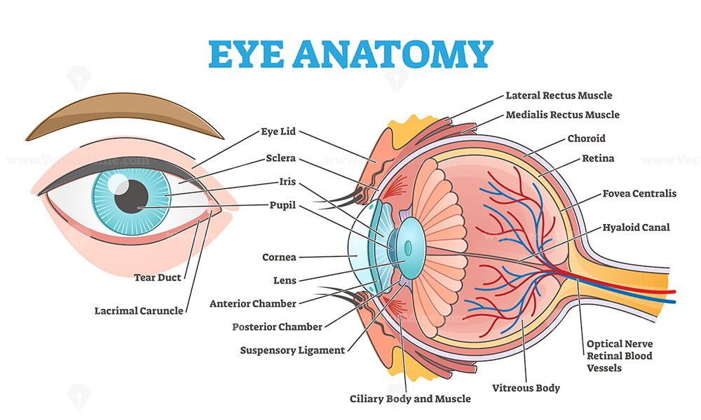
Eye Anatomy: Parts of the Eye and How We See Here is a tour of the eye starting from the outside, going in through the front and working to the back. Eye Anatomy: Parts of the Eye Outside the Eyeball. The eye sits in a protective bony socket called the orbit. Six extraocular muscles in the orbit are attached to the eye. These muscles move the eye up and down, side to side, and rotate the eye.

Eye diagram and labels
Structure and Functions of Human Eye with labelled Diagram - BYJUS The External Structure of an Eye. Sclera: It is a white visible portion. It is made up of dense connective tissue and protects the inner parts. Conjunctiva: It lines the sclera and is made up of stratified squamous epithelium. It keeps our eyes moist and clear and provides lubrication by secreting mucus and tears. human eye diagram with labels Draw A Labeled Diagram Of Human Eye Write The Functions Of Cornea, Iris, , eye draw diagram human labeled structure write iris labelled internal section retina lens cornea function functions eyeball pupil neat ball, Human Eye Structure: Eye Anatomy Explained - YouTube, , Eye Diagram: Label Quiz - PurposeGames.com Tournaments (37) AI Stream The more you play, the more accurate suggestions for you. Cities by Landmarks 11p Image Quiz. The worlds tallest buildings 9p Image Quiz. I spy on... 26p Image Quiz. The Western States 11p Image Quiz. Highscores (6 registered players) Member. Score.
Eye diagram and labels. The Eye Diagram: What is it and why is it used? The eye diagram is used primarily to look at digital signals for the purpose of recognizing the effects of distortion and finding its source. To demonstrate using a Tektronix MDO3104 oscilloscope, we connect the AFG output on the back panel to an analog input channel on the front panel and press AFG so a sine wave displays. Then we press Acquire. Diagram of the Eye - Lions Eye Institute The eye - one of the most complex organisms in the human body. It is made up of many different parts working in unison together. In order for the eye to work at its best, all parts must work well collectively. To understand the eye and its functions, it's important to understand how the eye works, see below diagrams for both the external ... 6,819 Human eye diagram Images, Stock Photos & Vectors | Shutterstock Find Human eye diagram stock images in HD and millions of other royalty-free stock photos, illustrations and vectors in the Shutterstock collection. Thousands of new, high-quality pictures added every day. Label Parts of the Human Eye - University of Dayton Parts of the Eye. Select the correct label for each part of the eye. The image is taken from above the left eye. Click on the Score button to see how you did. Incorrect answers will be marked in red. ...
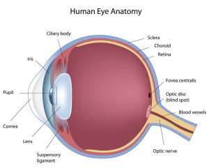
Labeled Eye Diagram | Science Trends The human eye is composed of many different parts that work together to interpret the world around us. What you want to interpret as a major part of the human eye is somewhat up to the individual, but in general there are seven parts of the human eye: the cornea, the pupil, the iris, the lens, the vitreous humor, the retina, and the sclera. Let's take a closer look at each of these ... Labelling the eye — Science Learning Hub Labelling the eye, Interactive, Add to collection, Use this interactive to label different parts of the human eye. Drag and drop the text labels onto the boxes next to the diagram. Selecting or hovering over a box will highlight each area in the diagram. Optic nerve, Lens, Schlera, Pupil, Vitrous humour, Iris, Cornea, Retina, Download Exercise, Eye Anatomy Diagram - EnchantedLearning.com Eye Anatomy. Our eyes are organs that let us see. Eyes detect both brightness and color. Having two eyes separated on our face enables us to have depth perception (the ability to see the world in three dimensions - 3D). How we see: A whole series of events happens in order for us to see something. First, light must reflect off an object. Anatomy of the eye: Quizzes and diagrams | Kenhub Try our crash course in eye anatomy. One of our favorite ways to get to grips with all of the parts of the eye is by utilizing labeled diagrams. On a diagram of the eye, we can see all of the relevant structures together on one image. This helps us to understand how each one is situated and related to the other. Labeled diagram of the eye,
Labeled Eye Diagram | Eye anatomy diagram, Eye anatomy, Diagram of the eye Eye Structure. Iridology. Simple eye diagram | Easy eye diagram | Labeled eye diagram We provide you Simple eye diagram and easy eye diagram from exam point of view. Also labeled eye diagram and anatomy of eye and human eye structure for better understanding. Human eye diagram and functions with diagram of human eye with labelling. Eye diagram labeled - Healthiack Now, let's study the parts of the eyes. Please click on the picture (s) to view larger version. Feel free to search healthiack.com for more details on this particular topic. Best viewed on 1280 x 768 px resolution in any modern browser. Eye diagram labeled 782, Eye diagram labeled 792, Eye diagram labeled 798, Eye diagram labeled 799, Eye diagram basics: Reading and applying eye diagrams - EDN Eye diagrams provide instant visual data that engineers can use to check the signal integrity of a design and uncover problems early in the design process. Used in conjunction with other measurements such as bit-error rate, an eye diagram can help a designer predict performance and identify possible sources of problems. Also see : Eye labeling Diagram | Quizlet a ring of muscle tissue that forms the colored portion of the eye around the pupil and controls the size of the pupil opening, Cornea, The clear tissue that covers the front of the eye, Posterior Compartment, filled with vitreous humor, Pupil, opening in the center of the iris, Susponsory Ligament, Allows the eye to move up and down,
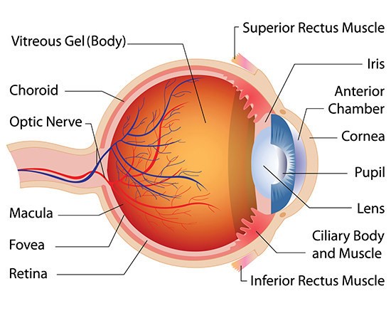
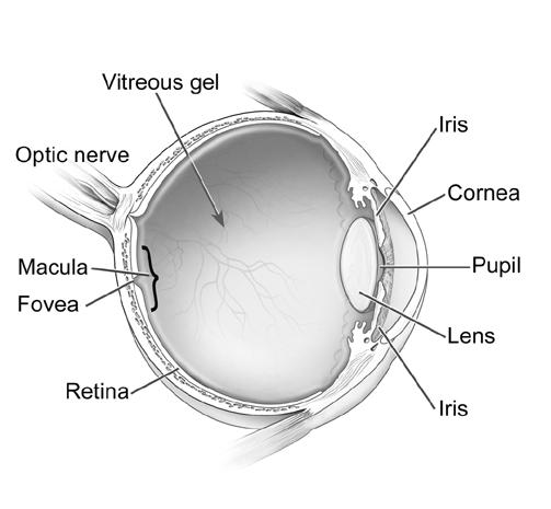
What Does the Eye Look Like? - Diagram of the Eye | Harvard Eye Associates Vitreous Gel: A thick, transparent liquid that fills the center of the eye. It is mostly water and gives the eye its form and shape. Our eyes are vital for seeing the world around us. Keep them healthy by maintaining regular vision exams. Contact Harvard Eye Associates at 949-951-2020 or harvardeye.com to schedule an appointment today.
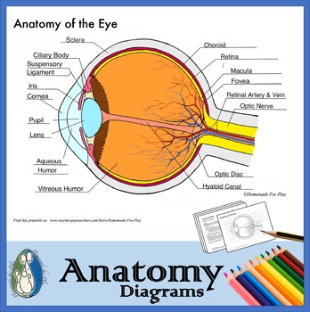
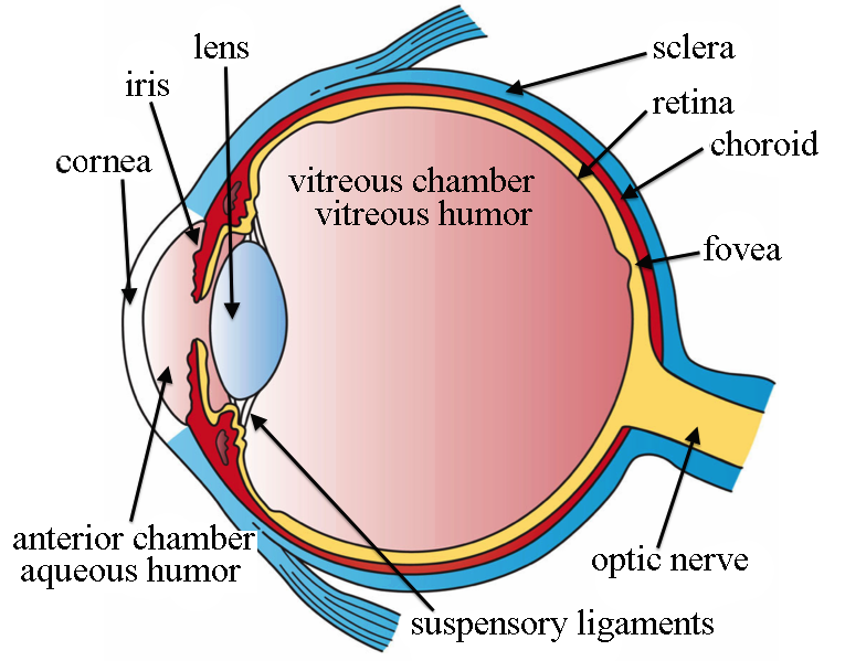
Eye Diagram With Labels and detailed description - BYJUS A brief description of the eye along with a well-labelled diagram is given below for reference. Well-Labelled Diagram of Eye, The anterior chamber of the eye is the space between the cornea and the iris and is filled with a lubricating fluid, aqueous humour. The vascular layer of the eye, known as the choroid contains the connective tissue.
Eye Diagram: Label Quiz - PurposeGames.com Tournaments (37) AI Stream The more you play, the more accurate suggestions for you. Cities by Landmarks 11p Image Quiz. The worlds tallest buildings 9p Image Quiz. I spy on... 26p Image Quiz. The Western States 11p Image Quiz. Highscores (6 registered players) Member. Score.
human eye diagram with labels Draw A Labeled Diagram Of Human Eye Write The Functions Of Cornea, Iris, , eye draw diagram human labeled structure write iris labelled internal section retina lens cornea function functions eyeball pupil neat ball, Human Eye Structure: Eye Anatomy Explained - YouTube, ,
Structure and Functions of Human Eye with labelled Diagram - BYJUS The External Structure of an Eye. Sclera: It is a white visible portion. It is made up of dense connective tissue and protects the inner parts. Conjunctiva: It lines the sclera and is made up of stratified squamous epithelium. It keeps our eyes moist and clear and provides lubrication by secreting mucus and tears.









![Cross sectional diagram of human eye [1]. | Download ...](https://www.researchgate.net/publication/276541864/figure/fig1/AS:612895498964992@1523137082339/Cross-sectional-diagram-of-human-eye-1.png)






Post a Comment for "43 eye diagram and labels"